Overview of Cell Death
Mechanisms and Types of Cell Death
Cells can die in a number of different manners, depending on the cellular context and triggering stimulus.
Types of Cell Death
Cell death mechanisms include:
- Apoptosis - programmed cell death that occurs during growth and development and can also occur in response to harmful environmental stimuli.
- Necrosis - can be a passive or an active, regulated process such as necroptosis or pyroptosis.
Different assays can be used to determine the mechanism of cell death or rule out a mechanism of cell death within a cellular population.
Apoptosis
Apoptosis is a highly regulated form of programmed cell death that occurs in multicellular organisms during development, throughout the lifespan, and in response to cellular stress. Apoptosis is mediated by a family of proteolytic enzymes called caspases. Other proteins, including proapoptotic and antiapoptotic proteins, also play important roles.
Dysregulation of apoptosis occurs in several disease states, including autoimmune disorders, neurodegenerative diseases, and cancer.
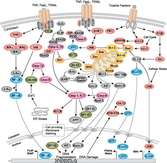
Apoptosis is a form of programmed cell death mediated by caspases and a host of other proteins.
Regulation of Apoptosis: Interactive Pathway >>
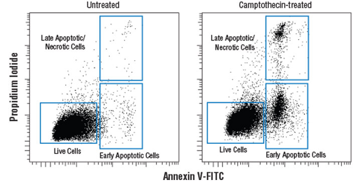
How to Measure Apoptosis
Apoptotic cells can be distinguished from viable cells by phenotypic changes and activity of certain proteins. Several different methods can be used to analyze and measure levels of apoptosis within a population. These assays include:
Assay
What is Measured
Detects changes that occur to the lipid bilayer early in apoptosis
Assay
TUNEL Kits
DNA fragmentation which is a hallmark of apoptosis
What is Measured
Assay
Caspase cleavage
What is Measured
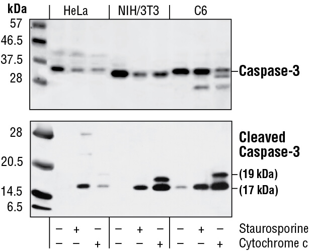
Cleavage of caspase-3, caspase activity assay, and cleavage of other caspases and PARP are frequently used readouts for apoptosis
Assay
Chromatin condensation
What is Measured
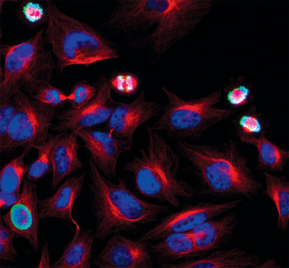
Detects apoptotic cells with condensed chromatin after staining with nuclear dyes, such as DAPI or Hoechst 33342
Assay
Cytochrome c release
What is Measured
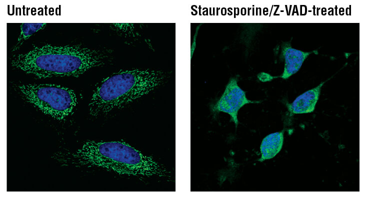
Translocation of cytochrome c from mitochondria to the cytoplasm is a hallmark feature of apoptotic cells
Assay
Mitochondrial membrane potential assay
What is Measured
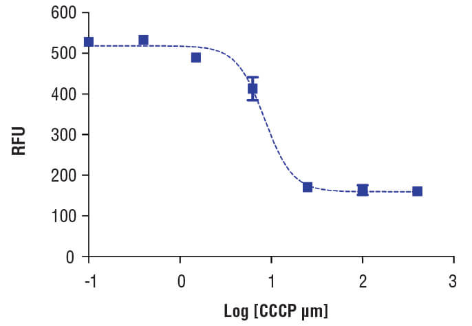
Depolarization and subsequent decrease of the mitochondrial membrane potential is a hallmark feature of apoptotic cells
Necrosis
Necrosis has been classically defined as an unprogrammed cell death that occurs following acute injury or infection or when apoptosis is inhibited and is characterized by cellular swelling and lysis. Necrotic cells release intracellular contents into the surrounding environment, which activates an inflammatory response to recruit phagocytes to clear dead cells. Uncontrolled, however, necrosis can cause severe tissue damage, such as gangrene.
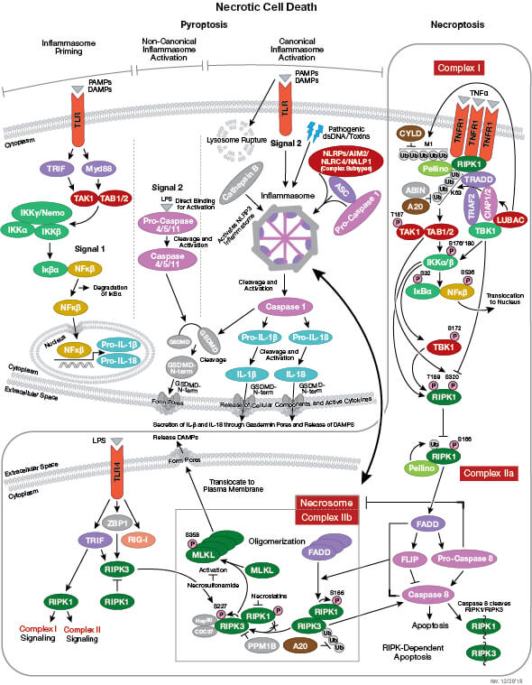
While it was previously thought that necrosis was passive and unprogrammed, recent data have uncovered different types of regulated necroptotic pathways.
Types of Regulated Necrosis
In addition to necrosis, other lytic cell death mechanisms include:
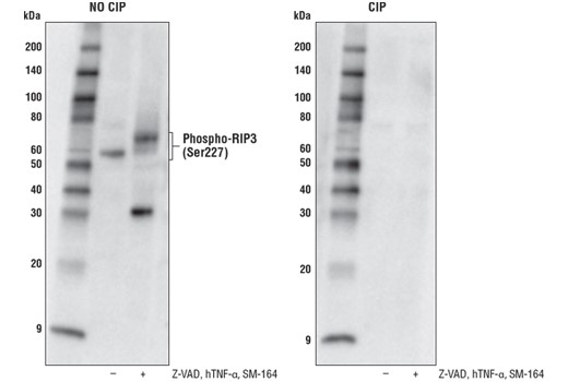
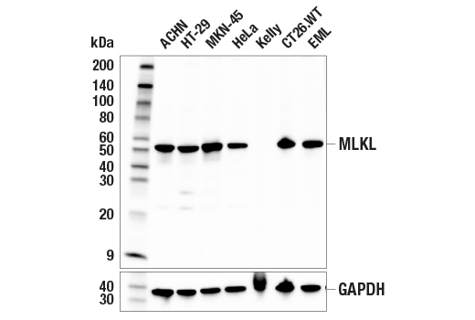
- Necroptosis - a programmed and regulated form of necrosis which requires RIP3 and MLKL and is activated by pro-inflammatory signaling as well as ischemic injury and viral infection.
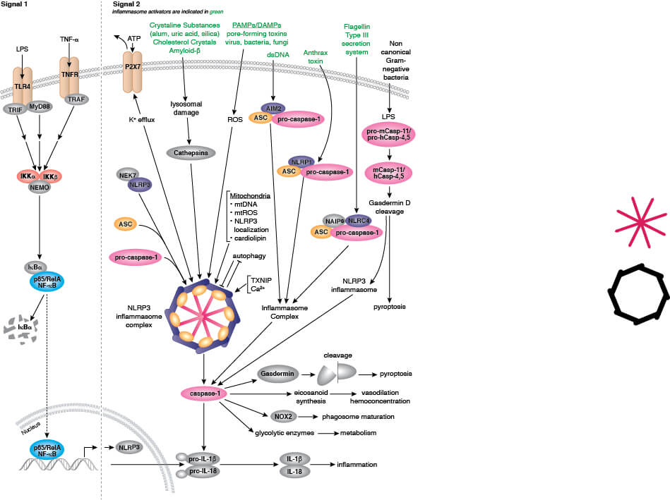
- Pyroptosis - a form of programmed lytic cell death that typically occurs in immune cells in response to microbial or viral infection and requires caspase-1 and gasdermin-D
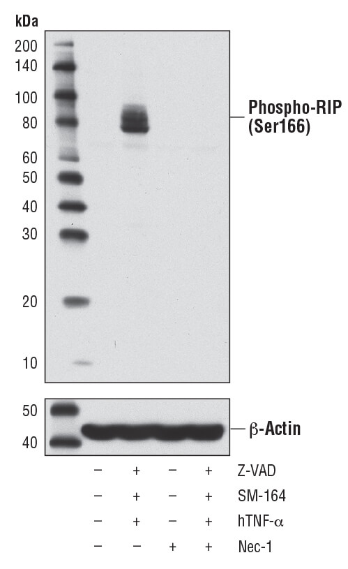
How to Measure Necroptosis:
Necroptosis Marker
Necroptosis Marker Description
Ser/Thr kinase that regulates inflammation and cell death
Necroptosis Marker
Necroptosis Marker Description
Ser/Thr kinase that is required for necroptosis
Necroptosis Marker
Necroptosis Marker Description
Activated RIP associates with RIP3 to trigger necroptosis
Necroptosis Marker
Necroptosis Marker Description
Activation of RIP3 leads to phosphorylation of MLKL
Necroptosis Marker
Necroptosis Marker Description
Downstream protein target of RIP3
Necroptosis Marker
Necroptosis Marker Description
Phosphorylation of MLKL leads to pore formation and is a marker for necroptotic cells
How to Measure Pyroptosis:
Pyroptosis Marker
Inflammasome formation
Pyroptosis Marker Description
Pyroptosis is characterized by the formation of the inflammasome; a marker for the inflammasome is NLRP3
Pyroptosis Marker
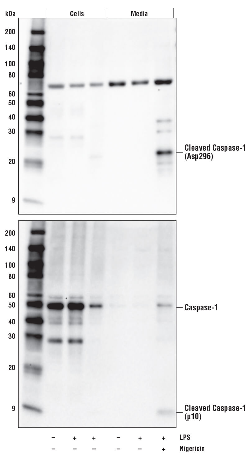
Caspase-1 activity
Pyroptosis Marker Description
Cleavage of caspase-1 is a marker for its activity. Activated caspase-1 cleaves IL-1β and gasdermin D
Pyroptosis Marker
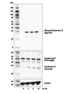
Gasdermin cleavage
Pyroptosis Marker Description
Cleavage of gasdermin-D occurs during pyroptosis lead to pore formation
How to Assess Necrosis:
Necrosis Marker
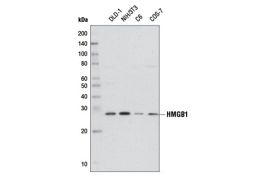
High mobility group protein B1 (HMGB1)
Necrosis Marker Description
Nuclear protein that is released into the extracellular environment during necrotic, but not apoptotic, cell death
Necrosis Marker
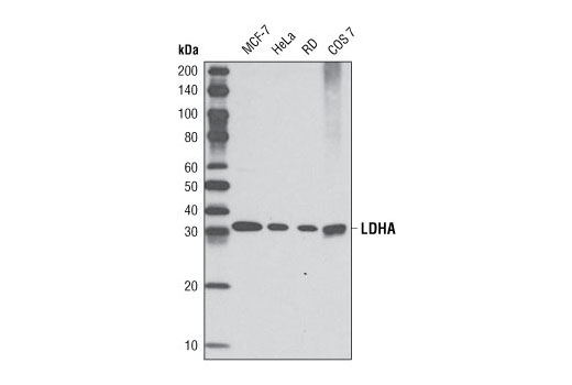
Lactate dehydrogenase A (LDHA)
Necrosis Marker Description
Cytosolic enzyme released into the extracellular space during necrotic cell death
Necrosis Marker
Necrosis Marker Description
Proinflammatory cytokine released during necrosis