

Immunology Research Center
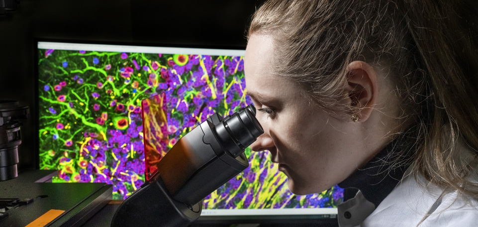
Fuel your immunology research with reliable and innovative tools.
Context matters, especially in immunology research. CST helps you keep up with the growing demand for spatial dimension insight across multiple modalities with rigorously tested, application-validated products that generate specific and reproducible high-content data, support from the scientists that developed the products, and a supply chain you can rely on.
See more and do more with:
- Antibody conjugates for immunohistochemistry (IHC)
- Antibody conjugates for flow cytometry
- CAR cell therapy research tools
- InTraSeq® Single Cell Analysis
- SignalStar® Multiplex IHC

Learn
Start with the basics.
Immunology research can range from immunophenotyping innate and adaptive immunity to unraveling intracellular and inflammatory signaling and characterizing the tumor microenvironment (TME).
Review immune mechanisms with these resources to expand your understanding of the biology before designing around it.

Phenotype and Functional Immune Cell Markers for Multiplex IHC
Additional blogs to build your inflammation knowledge.



Experiment
Match the method to the question.
Bring precision to your experimental design. Browse methods powered by application-validated antibodies, whether you are phenotyping, investigating spatial context, detecting secreted proteins, or investigating signaling responses.

Multiplex Solutions for Spatial Biology
Solutions for Antibody Pair-Based Immunoassay Development
Flow Cytometry Resource Center
Blog: Why Multiplex with Chimeric Antibodies?
Reproducible, specific solutions to advance your research.
Spatial Profiling with SignalStar Multiplex IHC

Customizable, flexible, and highly-validated antibody panels for your spatial biology research. Amplify multiple targets simultaneously in FFPE tissue with high sensitivity and specificity–and generate up to 8 targets in just 2 days.
InTraSeq Single Cell Analysis

Conjugate antibody cocktails, assay kits, and conjugates to investigate signaling pathways with the transcriptome at the single-cell level. Simultaneously quantify mRNA expression alongside intracellular and cell surface proteins in thousands of cells with InTraSeq (Intracellular protein and Transcriptomic Sequencing).
Immunohistochemistry Solutions

IHC-validated antibodies enable the detection and analysis of protein expression while maintaining the composition, cellular characteristics, and structure of the native tissue. Identify immune cell subsets, assess the tumor microenvironment, diagnose immune-mediated disorders, and more.
Resources to optimize your experiment design.

Poster Video
InTraSeq Single Cell Analysis: A Multimodal Single-Cell Method for Simultaneous Measurement of RNA and Proteins to Uncover New Single-Cell Biology

Guide
Antibody Staining Guide for Flow Cytometry

Blog
Antibody Compatibility Trifecta: Choosing a Multiplexing Strategy for Reliable Spatial Biology Insights
Services to support your research.

Customized and Carrier-Free Formulations
Conjugation-ready antibody formulations to match your assay platform.

Custom Antibody Conjugation & Labeling Services
Antibody conjugates to match your experimental design.

Bulk Quantities and Lot Reservations
Consistent supply of antibodies and reagents to future-proof your assay.

Characterize
Immune Cell Profiling with Uncompromising Precision.
Your data will only hold up if your studies are performed in the correct cell type in the appropriate activation state–meaning T cells should not be undergoing exhaustion when examining markers for robust effector function. Additionally, it is important to consider the phosphorylation status or subcellular translocation of relevant proteins when examining specific immune cascades.
Ensure your experiments are performed in the correct model with highly specific, validated antibodies against key immunological markers to confidently characterize immune cell populations and pathways.

Phenotype and Functional Immune Cell Markers for Multiplex IHC
Blog: Immunophenotyping using Flow Cytometry or IHC
Confident characterization with human & mouse immune cell marker guides.
Know your cell. Quickly identify key human and mouse immune cell markers and relevant CST® antibody products for your IHC and flow cytometry assays.

Cell Type Markers

Profiling tumor-infiltrating immune cells
Tumor-infiltrating immune cells are more than just a phenotype. This blog reviews both the cell types present and the functions they carry out.
Identifying lymphoid cells
Lymphoid cells include T cells, B cells, and NK cells. This blog walks you through what defines the lymphoid lineage and how each population is identified.
Identifying myeloid cells
Myeloid cells include monocytes, macrophages, neutrophils, and dendritic cells. This blog covers what defines the myeloid lineage and how each type is identified.

Discover
Map Your Research: Immunology Pathway Diagrams.
Use this curated collection of pathways and diagrams to help you examine immunological mechanisms. These meticulously researched and designed pathway diagrams represent evidence-based signaling information and can help you find the best products to fuel your research.

Immuno-oncology Signaling Pathways Poster
CAR Signaling Networks Interactive Pathway
View the Full Library of Signaling Diagrams

Translate
Connecting science to drug discovery.
Leverage the experience of CST scientists to streamline your drug discovery and accelerate therapeutic development. Keep up with project timelines with:
- Reliable, application-validated antibodies guaranteed to work
- Innovative tools, like SignalStar and InTraSeq, to see more with less
- Custom solutions and services to take the research, testing, and validation off your plate.
- Guaranteed supply continuity
Maximize Drug Discovery Insights–at Scale
Solutions for Cell Therapy Manufacturing and QC
High-Content Imaging to Drive Your Drug Discovery Forward
Solutions for Antibody Pair-Based Immunoassay Development
Chimeric Antibody Receptor (CAR) Solutions for your Drug Discovery.
CAR-Engineered Cell Characterization Solutions
Streamline your CAR-based cell therapy development. Detect, analyze, quantitate, and purify CAR-engineered cells faster and easier with robust, selective, and sensitive CAR linker antibodies that remove the need for an anti-idiotype antibody.
Deciphering CAR-T Cells: Exploring Functional Mechanisms to Drive Next-Generation Immunotherapy
Learn about the role different T cell populations play in normal immune function and disease, and how these breakthroughs can improve CAR-T cell immunotherapy effectiveness in treating diseases.
Immunotherapy and CAR-T Cells
Watch this video series designed to describe how CAR-T therapies are designed and implemented, and review challenges researchers developing these therapies face.
Analytical Tools to Evaluate CAR-T Cell Signaling & Activation
Learn how the precision of Simple Western, a capillary-based immunoassay, and CAR solutions from CST can work together to streamline the workflow to engineer and characterize CAR-T cell signaling and activation status.