

TGF-β Signaling
Access the full library of downloadable pathway diagrams, along with recommended products for each signaling pathway.
2026 © Cell Signaling Technology. All Rights Reserved.
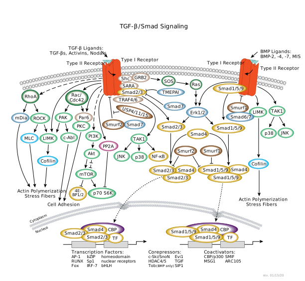
Transforming growth factor-β (TGF-β) superfamily signaling plays a critical role in the regulation of cell growth, differentiation, and development in a wide range of biological systems. In general, signaling is initiated with ligand-induced oligomerization of serine/threonine receptor kinases and phosphorylation of the cytoplasmic signaling molecules Smad2 and Smad3 for the TGF-β/activin pathway, or Smad1/5/9 for the bone morphogenetic protein (BMP) pathway. Carboxy-terminal phosphorylation of Smads by activated receptors results in their partnering with the common signaling transducer Smad4, and translocation to the nucleus. Activated Smads regulate diverse biological effects by partnering with transcription factors resulting in cell-state specific modulation of transcription. Inhibitory Smads, i.e. Smad6 and Smad7 antagonize activation of R-Smads. The expression of inhibitory Smads (I-Smads) 6 and 7 is induced by both activin/TGF-β and BMP signaling as part of a negative feedback loop. The stability of TGF-β family receptors and/or Smads are regulated by Smurf E3 ubiquitin ligases and USP4/11/15 deubiquitinases. TGF-β/activin and BMP pathways are modulated by MAPK signaling at a number of levels. Moreover, in certain contexts, TGF-β signaling can also affect Smad-independent pathways, including Erk, SAPK/JNK, and p38 MAPK pathways. Rho GTPase (RhoA) activates downstream target proteins, such as mDia and ROCK, to prompt rearrangement of the cytoskeletal elements associated with cell spreading, cell growth regulation, and cytokinesis. Cdc42/Rac regulates cell adhesion through downstream effector kinases PAK, PKC, and c-Abl following TGF-β activation.
Selected Reviews:
- Horbelt D, Denkis A, Knaus P (2012) A portrait of Transforming Growth Factor β superfamily signalling: Background matters. Int. J. Biochem. Cell Biol. 44(3), 469–74.
- Ikushima H, Miyazono K (2010) TGFbeta signalling: a complex web in cancer progression. Nat. Rev. Cancer 10(6), 415–24.
- Kitisin K, Saha T, Blake T, Golestaneh N, Deng M, Kim C, Tang Y, Shetty K, Mishra B, Mishra L (2007) Tgf-Beta signaling in development. Sci. STKE 2007(399), cm1.
- Meulmeester E, Ten Dijke P (2011) The dynamic roles of TGF-β in cancer. J. Pathol. 223(2), 205–18.
- Schmierer B, Hill CS (2007) TGFbeta-SMAD signal transduction: molecular specificity and functional flexibility. Nat. Rev. Mol. Cell Biol. 8(12), 970–82.
- Xiao YT, Xiang LX, Shao JZ (2007) Bone morphogenetic protein. Biochem. Biophys. Res. Commun. 362(3), 550–3.
- Zhang L, Zhou F, García de Vinuesa A, de Kruijf EM, Mesker WE, Hui L, Drabsch Y, Li Y, Bauer A, Rousseau A, Sheppard KA, Mickanin C, Kuppen PJ, Lu CX, Ten Dijke P (2013) TRAF4 promotes TGF-β receptor signaling and drives breast cancer metastasis. Mol. Cell 51(5), 559–72.
We would like to thank Prof. Peter ten Dijke, Leiden University Medical Center, Leiden, The Netherlands, for contributing to this diagram.
created January 2003
revised June 2014